Meet Dr. Yana Zorina
Neurobiologist and Artist
Dr. Yana Zorina is a neurobiologist and Senior Research Scientist at Memorial Sloan Kettering Cancer Center (MSKCC), where she helps test a collection of chemical compounds for various cellular responses using biochemical and image-based assays. Working in cell biology, she is often exposed to microscopy techniques and the beautifully complex structures that make up the mammalian brain. Also as the artist behind “NeuroBead”, she draws from her neuroscience knowledge to transform confocal microscopy images into precise 3D beadwork that encourages a wider audience to appreciate the beauty of scientific research.
STEM to the Sky
Jul 9, 2021
- Can you explain what your work entails at MSKCC?
- How exactly does microscopy work?
- When did you become interested in neuroscience and research?
- How did your experiences in graduate school prepare you for your current role?
- What was your motivation behind combining neuroscience and art to start NeuroBead?
- How do you find time to pursue both research and NeuroBead?
- How do we help the public appreciate the beauty of science and why is it important?
- What would a typical day for you look like working at MSKCC?
- What is the most rewarding aspect of your work?
- Is there anything that surprised you about either your role or your field when you first started?
- What is the most challenging aspect of your career?
- What skills aside from the technical skills are important for a role like yours?
- Do you have any advice for combining an interest in science with another passion?
- What would you say to a student who is interested in pursuing neuroscience/cell biology in general?
Can you explain what your work entails at MSKCC?
I was initially trained during my undergrad and graduate studies as a neuroscientist, but when you get into that field, you also get highly exposed to cell biology. Over the past four years, I actually switched my job a little bit, to work in a cancer center where we take all sorts of different cells, treat them with large collections of compounds, and then look at phenotypic assays. This usually involves microscopy, which uses many fluorescent tags and stains to see cellular responses to different compounds and drugs.
Rather than the traditional approach of using logic to think “If this is my signaling pathway, then maybe I can target A or B here to get the response that I want”, high throughput screening tests thousands of compounds to see which ones give you the desired response.

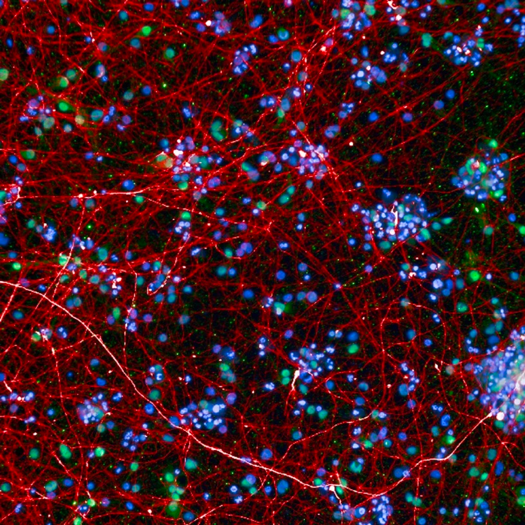
Neurons under confocal microscope (Credit: Dr. Yana Zorina)
How exactly does microscopy work?
I’ve loved microscopy for pretty much all of my professional life. I started using it in graduate school, and I've used it throughout my postdoctoral training up until now.
Cells consist of proteins, and you can look for your protein of interest to see where it is expressed and localized in the cell. For that, you can use antibodies that can bind to the protein, and you can stain those antibodies with fluorescent tags in all kinds of different colors. Then, you get images of the cells that show you where your protein of interest is localized. Once you capture these images, you can upload them into software to quantify all sorts of different parameters such as shape, intensity, number of cells, cell survival/death, and just about anything you can think of.
When did you become interested in neuroscience and research?
I got interested in biology and neuroscience probably around 6th or 7th grade. I was determined to go into something biology related, but at the time I was ignorant of the fact that you can actually go into science. I thought that if I was interested in biology, then medical school is the way to go.
In high school, I participated in a research program where I worked in a lab. I continued working in a different lab during college, where I majored in neuroscience. I went into pre-med as the default choice, but that didn’t mean that I had to go to medical school; I could actually go into biology and research.
I remember that while I had this idea that I would go to medical school, I realized that I didn’t really want to. I went to a career advisor in my college and I told her that I don't think that medical school was for me.
She said a great thing. There are three stages of science: researchers who discover the knowledge, teachers who spread the knowledge, and then doctors who apply it. Each one of these stages is equally important. I wanted to be at the beginning of the process.


(Credit: Dr. Yana Zorina)
How did your experiences in graduate school prepare you for your current role?
When I was going into graduate school, I was very narrow-minded in a sense. I selected a graduate school based on a specific lab that I wanted to work in, which then turned out that they couldn't take me. During the first year and a half of graduate school, you do two or three rotations, where you try out different labs for two or three months each. That's how you decide which one to go into for your training.
I thought I was going to do all of my rotations in the Department of Neuroscience, until somebody recommended looking into the Department of Pharmacology. I was pleasantly surprised with how much neuroscience was going on there as well. Being exposed to basic cell biology and signaling pathways that can be applied in essentially any field gave me a real advantage.
Right now, I'm working at a cancer center, but during graduate school, I studied signaling pathways that would potentially lead to regeneration of nerve cells. A lot of the same genes are implicated in cancer.
What was your motivation behind combining neuroscience and art to start NeuroBead?
I’ve always done a lot of art, and going into art definitely did cross my mind a couple times in school. But, I kept telling myself that I was too busy with my work with family, and so forth.
In 2016, I just had this acute sense that art is really a part of me and is something I need to bring back into my life. As I started thinking about what I would like to do and what my art should be about, I thought “why not join both sides of my brain?”.
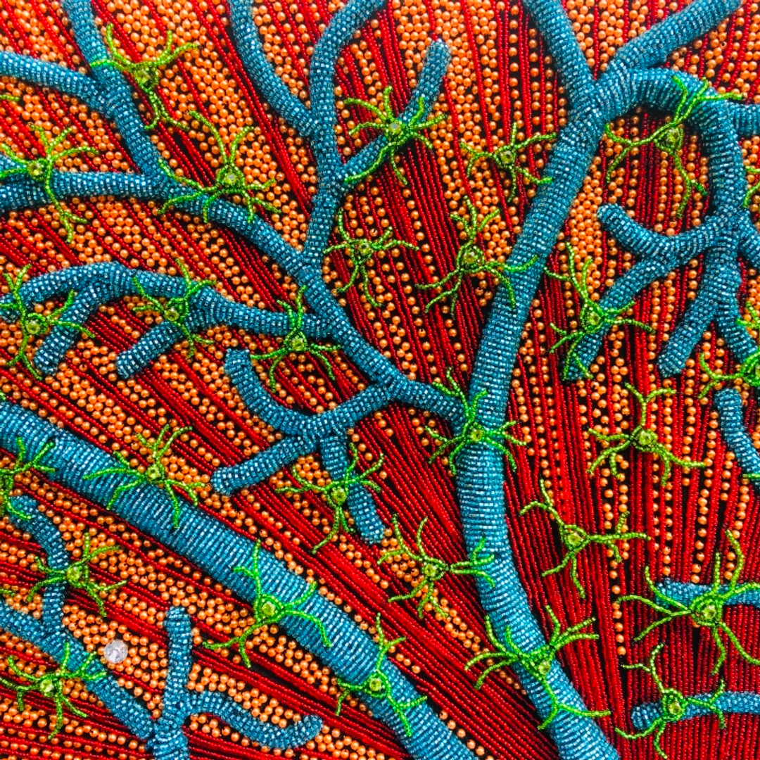
There is a method called confocal microscopy, which uses fluorescent channels. The point of it is that the microscope is able to focus on a very thin optical section, so that you know exactly what is at that level.
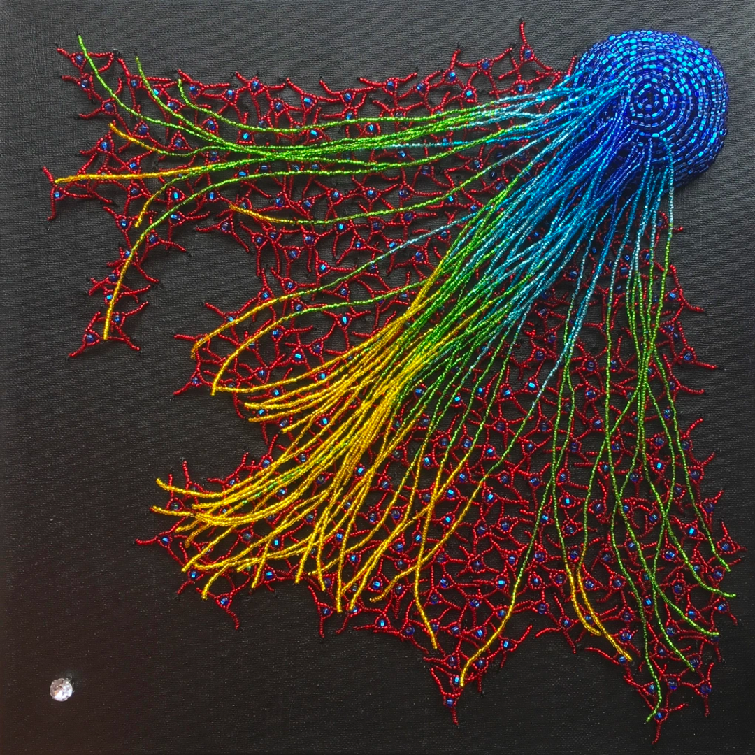
If you take confocal microscopy images, what would happen if you turn them on their head and recreate them in 3D? That's what I wanted to do to get a better idea of what whole cells or tissues may look like. Thinking of fluorescence microscopy, I immediately thought back to my experience as a teenager making beadwork and thought, “Well, beads provide a great analogy to pixels!”
"Finding Your Self"
"Attraction"

"Fragile Memory"
(Credit: Dr. Yana Zorina)
How do you find time to pursue both research and NeuroBead?
I jokingly refer to myself as a scientist by day and an artist by night. I have a relatively stable 9am -5pm schedule. After work, I come back home and spend time with my family. Once the kids are out, I run to my art table, and that's really the “me-time” that gives me a sense of flow. It allows the left hemisphere of my brain as a scientist to relax. Letting go of the constant logical thinking that I have to do during the day helps me tune into my intuition a bit more.
How do we help the public appreciate the beauty of science and why is it important?
Years ago, there were many political articles saying that the government is throwing tax money into ridiculous research that no one cares about. The example they gave was an image of a shrimp placed on a treadmill. There was this uproar, saying, “Where is our money going? Do they have nothing better to do with millions of taxpayers’ dollars? How ridiculous is this?”
A month or two later, the graduate student who did that work actually wrote his own article where he explained, “I am the one who placed the shrimp on the treadmill.” He said that it did not cost millions of dollars and that he constructed it himself for $50 out of his own pocket. As a matter of fact, the reason that he did this was because he wanted to see how marine animals can fight infection. For that, the animals need to be placed under physical stress.
The take home message is that to understand why science is important, people outside of the so-called “ivory tower” of academia should be able to understand and interpret the science that goes on behind closed doors. There is greater movement now with science communication, so that people don't think that what happens in a research lab is ridiculous.
Art provides a great channel for starting these conversations and makes science a bit more digestible to the general public who do not deal with it everyday.
"To understand why science is so important, people outside of the so-called "ivory tower" of academia should be able to understand and interpret the science that goes on behind closed doors"
Dr. Yana Zorina
What would a typical day for you look like working at MSKCC?
I'm working at a core facility, so my week is typically sprinkled with a lot of meetings with either new clients or reporting on the status of existing projects. In between all of that, I spend time culturing cells in the lab. I grow them up to a desired quantity and arrange them into plates that have hundreds of wells. Then, I perform the experiments. I treat the cells and fix them in place almost like fixing tissue for pathology.
There's a lot of imaging and image analysis, and then the rest of the time is spent crunching the numbers and seeing which compounds may give you a desired response. Then, I go back to the literature to see if it makes sense and check if somebody has found something similar in that disease area. Finally, I put together PowerPoint slides to present it to other people.
What is the most rewarding aspect of your work?
I really like image analysis. It requires a lot of attention to detail. It’s really rewarding and almost counterintuitive when you can take pictures and turn them into numbers that actually mean something. I really like looking at individual cellular responses and how very minor cellular effects can actually have a great impact on a larger disease area.
For example, something that I came across relatively recently is a technique called Cell Painting, where you apply five to six dyes in different fluorescent colors. Rather than looking for a specific response that you would be interested in, you look at the overall cell state. Based on that, you can make an educated guess of what is going on in a cell and what processes are worth looking into and researching further. It's just such a fascinating way to really narrow down what may be happening in an organism and then translate it into new disease models.
Over the past few years and probably the next decade or so, a lot of exciting new methods are coming out including spheroid and organoid cultures. Rather than the traditional method of culturing individual cells on a flat surface, you can now actually create tiny 3D organs, that you can put into a dish, of different types of tissues that you're interested in. This technique allows you to minimize both studies in animal models, as well as potentially reduce the amount of work that needs to be in clinical trials.
Is there anything that surprised you about either your role or your field when you first started?
One of the things that really surprised me is how many common pathways there are in all sorts of different tissues. It almost doesn't matter what tissue or type of disease you're working on. One way or another, you're going to come across the usual suspects. You can probably count how many central genes there are on your fingers.
It's mind boggling in a way that you feel right at home when you see a familiar gene name, but then you wonder how it has completely different roles in different contexts. For example, a lot of the proteins that I worked on in regeneration of the central nervous system are oncogenes that are implicated in cancer. While we were trying to activate them, oncologists are actually trying to suppress them. Everything is good in moderation. So you need to find how to fine tune everything, so that you have just enough of it, but not too much of a good thing that could throw the balance in the opposite direction.
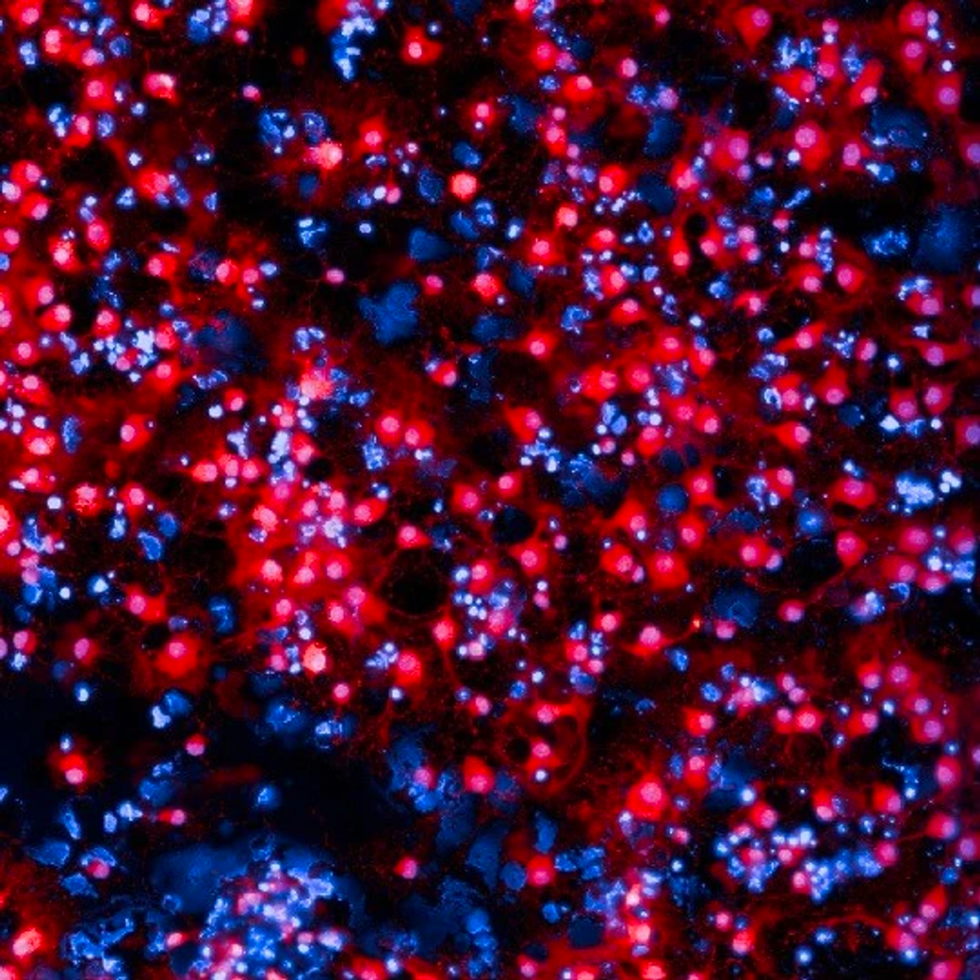
Oligodendrocytes under confocal microscope (Credit: Dr. Yana Zorina)
What is the most challenging aspect of your career?
In one of the projects that I worked on for a long time, we got a lot of different types of information from different experiments. It's both challenging and rewarding to be able to take those seemingly unrelated bits and pieces and fit into one coherent picture. It takes a lot of time and thinking; you need to figure out what questions to ask next in order to plug in the holes that you're missing.
What skills aside from the technical skills are important for a role like yours?
Going back to communication, you need to be able to explain to people what you do and why it matters. One phrase that gets thrown around a lot is that “if you really know what you're doing, you should be able to explain it to a five year old”. Also, you should make sure to be open to collaboration and other ideas. With different signaling mechanisms that can be applied in many fields, for example, you need to be able to learn from other areas that you may not be particularly familiar with. Maintain an open mind to see how you can apply what you have learned in your research.
Do you have any advice for combining an interest in science with another passion?
Art was a hobby for me that I decided to take to the next level. It's really important to retain your interests and not let them fall by the wayside. Whether it's art or a musical instrument or a type of sport, it allows science to not completely consume you. There are other multifaceted sides to you, and that’s actually what makes you interesting to other people, including those within the scientific community. It's important to nurture and develop those hobbies, whether you take them to the next serious level or not.
What would you say to a student who is interested in pursuing neuroscience/cell biology in general?
It's definitely great to go into biology. You need to learn a little bit of everything to see what actually interests you. I would say go in knowing what you're interested in, but at the same time, be open minded to what you might stumble upon. You never know what might pique your interest on another day.
It's important to realize that what you learn in one area may become unexpectedly relevant in another one. You never know how different types of knowledge may come in handy. In general, it is important to have a plan to know what kind of milestones you need to reach in order to get to the target that you have set for yourself. But at the same time, be open minded to what may come your way.
For those interested in neuroscience, there is an annual conference called Society for Neuroscience. The spectrum of neuroscience goes from molecular biology all the way up through behavior. I don't think that any other field has such a wide spectrum!
Learn More:
- NeuroBead Website
- Instagram: @neurobead_boutique
- Twitter: @YZorina
- Facebook: NeuroBead
- Etsy: NeuroBead
- Society for Neuroscience